
By Team INVC | INVC NEWS
STANFORD, California, United States | September 17, 2026 —
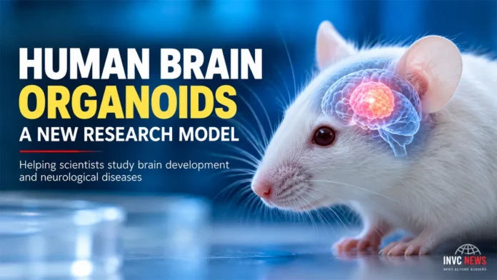
Scientists have developed a new way to study human brain development by growing laboratory-made human cortical tissue inside specially engineered mice.
The research uses human brain organoids — small, three-dimensional clusters of neural cells grown from human stem cells — and places them into mice whose cerebral cortex did not develop normally.
Researchers call the approach xenocortication.
The experiment does not create a mouse with a human brain. Nor does the research show that the animals developed human intelligence or consciousness.
Instead, scientists created a living experimental environment in which human neurons could grow, mature, form connections and respond to injury.
That could eventually help researchers understand brain development and neurological disease more accurately.
What Are Human Brain Organoids?
Human brain organoids are tiny collections of neural cells grown in a laboratory from stem cells.
Scientists can guide these stem cells to develop characteristics found in particular regions of the human brain.
In this study, researchers used human cortical organoids, which resemble aspects of the developing cerebral cortex.
The cerebral cortex plays an important role in functions including perception, language, attention, decision-making and cognition.
However, an organoid is not a miniature complete human brain.
It lacks many of the structures, connections and biological systems found in a living human brain.
That limitation is one reason researchers wanted to place the tissue inside a living organism.
Why Scientists Used Specially Engineered Mice
Previous experiments have transplanted human neural organoids into rodents.
However, normal rodent brains leave relatively little physical space for human tissue to grow.
Existing mouse or rat neurons also compete with transplanted human neurons while connections form.
The Stanford-led research team took a different approach.
Researchers used mice genetically engineered so that much of their neocortex and hippocampus did not develop normally.
Scientists describe these animals as apallial mice.
The missing cortical tissue created space where transplanted human organoids could expand and connect with the remaining mouse nervous system.
Researchers transplanted the organoids into mice when the animals were only a few days old.
Human Tissue Survived and Grew
The transplanted human tissue survived in most of the animals examined.
Researchers reported graft survival in about 86% of transplanted mice assessed through MRI.
The human-derived tissue then expanded significantly.
Between two and three months after transplantation, graft volume increased by roughly 4.7 times in the animals examined.
In a smaller group assessed at three months, human-derived organoid tissue represented, on average, more than 90% of the combined cortical tissue volume.
That does not mean more than 90% of the animals’ entire brains became human.
The figure refers specifically to the relevant cortical tissue measured in those xenocortical mice.
This distinction matters because dramatic numbers can easily create a misleading impression when removed from their scientific context.
Human Neurons Connected With the Mouse Nervous System
Growth alone was not the study’s most important finding.
Human neurons also integrated with the remaining mouse nervous system.
Researchers observed nerve fibres extending from the human graft into other parts of the brain.
Electrical recordings and calcium imaging also detected organized activity within the transplanted tissue.
The findings suggest that human neurons did not simply remain as isolated clusters.
Instead, they became part of functioning neural circuits inside the animals.
This gives researchers a new environment for studying how human neurons develop when they receive blood supply, sensory input and signals from a living nervous system.
Did the Mice Develop Human Intelligence?
No evidence from the study shows that the mice developed human intelligence, a human personality or human-like consciousness.
This is one of the most important points for readers to understand.
Researchers reported that the xenocortical mice generally retained locomotor abilities comparable with other mice, although they observed selective differences in limb coordination and patterns of spontaneous behaviour.
Those findings require scientific interpretation.
They do not demonstrate that the animals acquired human cognition.
Headlines suggesting that scientists created “human-brained mice” would therefore oversimplify the research.
Why This Research Could Be Useful
Scientists face a major challenge when studying diseases of the human brain.
Living human brain tissue is difficult to access for obvious medical and ethical reasons.
Researchers consequently rely on laboratory cell cultures, organoids, donated tissue, imaging and animal models.
Each method has limitations.
Many neurological and developmental conditions involve complex networks of human neurons that ordinary laboratory mice cannot reproduce.
Human brain organoids offer another option, but tissue growing inside a laboratory dish cannot fully recreate the environment of a living nervous system.
Xenocortication attempts to bridge that gap.
It gives human-derived neurons room to mature and interact with a living biological system.
Researchers Tested Oxygen Deprivation
The scientists also tested whether the model could help them study brain injury.
They exposed xenocortical mice to a period of very low oxygen.
The human-derived cortical tissue showed a notable biological response to oxygen deprivation.
Researchers detected signs of injury in the human graft that were much less apparent in nearby mouse tissue.
That difference could eventually help scientists investigate why the developing human brain may be particularly vulnerable to oxygen deprivation.
Such research could contribute to a better understanding of conditions associated with brain injury around pregnancy or birth.
However, this experiment does not provide a treatment.
It establishes a research platform that scientists may use to study disease mechanisms and potential therapies.
A Rare Human-Associated Neuron Also Appeared
The researchers identified von Economo neurons, or VENs, within the human-derived tissue.
These unusual neurons are relatively rare and occur in certain brain regions associated with social awareness and decision-making.
Scientists have found them in humans and some other large-brained social animals.
Researchers are particularly interested in VENs because changes involving these cells have been associated with conditions including some forms of frontotemporal dementia.
Their appearance in xenocortical tissue may give scientists another way to investigate cell types that have been difficult to study in laboratory models.
Could This Help Researchers Study Neurological Disorders?
Potentially, but substantial work remains.
Researchers could eventually grow organoids from cells carrying genetic changes linked with particular neurological or neurodevelopmental conditions.
They could then observe how those human neurons mature and function inside a living neural network.
The model might also help scientists study how human neurons respond to experimental medicines.
Possible areas of future investigation include developmental brain disorders, neurological injury and conditions involving abnormal neural circuits.
Researchers must still establish how accurately findings from xenocortical mice reflect what happens inside a human brain.
No animal or organoid model can perfectly reproduce the complexity of a person.
Ethical Oversight Will Remain Important
Research involving human neural tissue and animals requires careful ethical oversight.
The Stanford team said the human cells used to create the organoids came from donors who had consented to their cells being transplanted into animals.
As human neural models become more sophisticated, scientists will need to continue examining animal welfare, experimental boundaries and the biological capabilities of increasingly complex organoids.
Clear communication will also matter.
Scientists and news organizations should distinguish between human neural tissue growing inside an animal and claims about human consciousness or identity that the evidence does not support.
What Does Xenocortication Mean?
The word combines the idea of cross-species transplantation with the cerebral cortex.
In practical terms, xenocortication means placing human-derived cortical organoids into an animal model where they can grow extensively and integrate with the host nervous system.
The approach gives researchers access to living human neurons while retaining the experimental advantages of an animal model.
That combination is what makes the technique scientifically interesting.
Is This a Medical Treatment?
No.
Xenocortication is currently an experimental research method.
Doctors are not transplanting these organoids into patients, and the study does not establish a new treatment for autism, epilepsy, cerebral palsy, schizophrenia, dementia or any other neurological condition.
Its immediate value lies in research.
Scientists hope the model will help them understand how human neural cells develop, how diseases disrupt them and how potential therapies affect them.
Turning discoveries from laboratory models into safe human treatments usually requires years of additional investigation.
Why the Study Matters
The human brain remains one of the most difficult biological systems to study.
Traditional animal models have transformed neuroscience, but important differences exist between rodent and human brain development.
Laboratory-grown organoids have helped close some of that gap.
Xenocortication takes the approach further by allowing human-derived cortical tissue to mature inside a living nervous system and form extensive connections.
The research therefore offers scientists a potentially valuable new model for exploring human neurodevelopment, brain injury and neurological disease.
Its significance does not come from creating an animal with a “human mind.”
It comes from giving researchers a new way to observe living human neural cells in an environment that laboratory dishes cannot reproduce.
As the technique develops, its scientific value will depend on rigorous experiments, careful interpretation and continued ethical oversight.
Official Research Sources
Nature — Peer-Reviewed Research Paper
Read the official Nature research paper
Stanford Medicine — Official Research Report
Read the official Stanford Medicine report
Readers who want to examine the original scientific findings should use the Nature paper as the primary research source. Stanford Medicine provides an accessible institutional explanation of the study, its methodology and its potential scientific applications.